
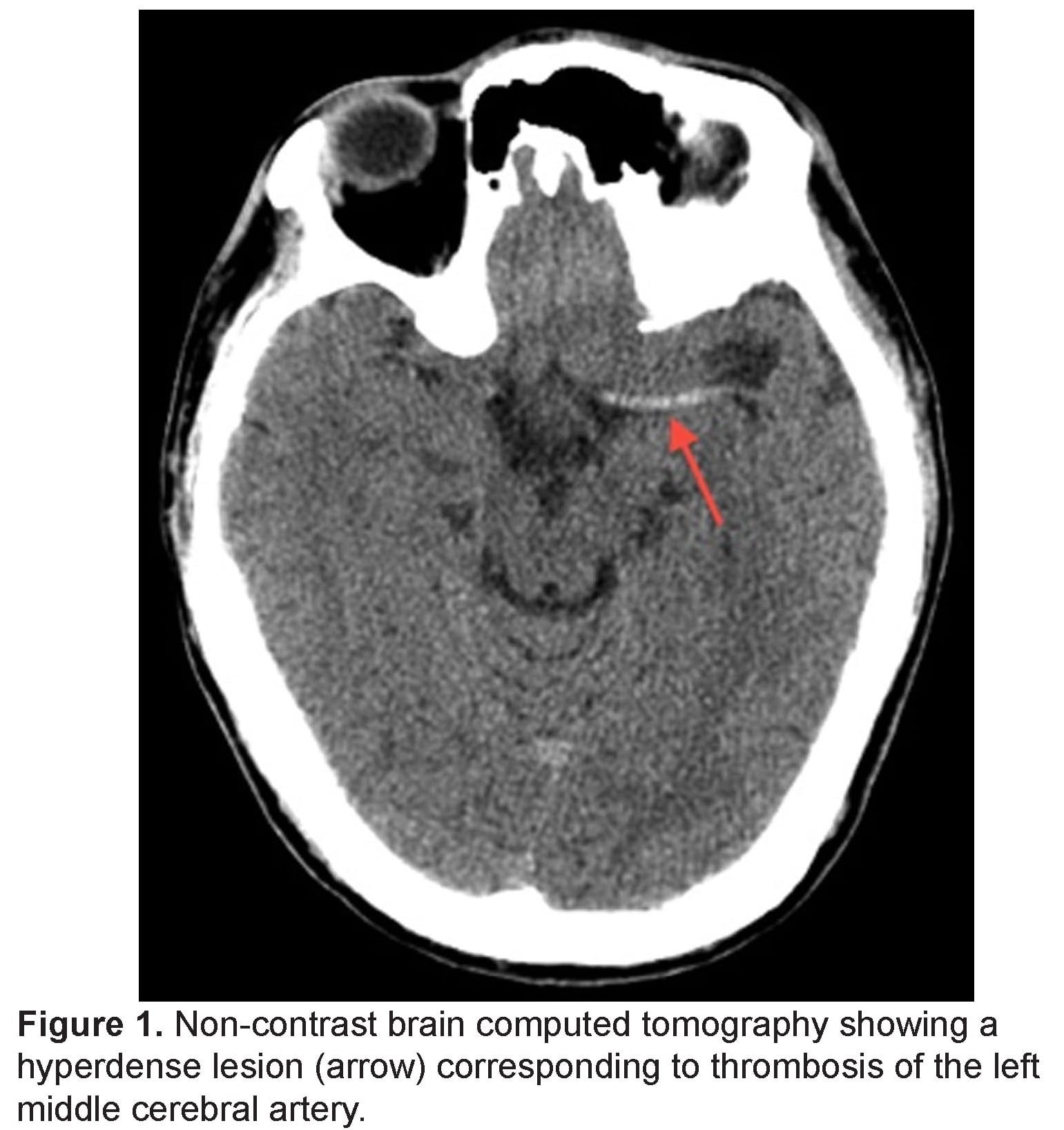
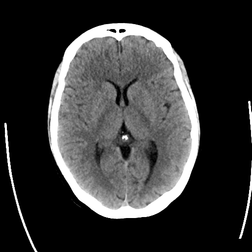
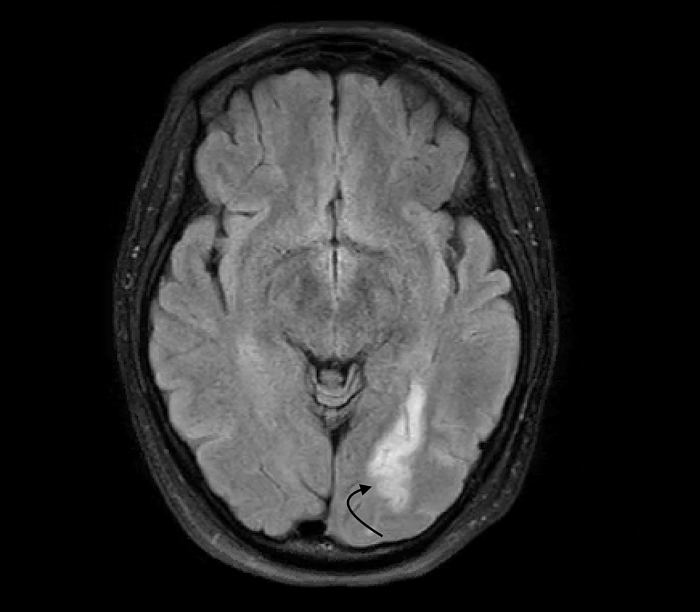
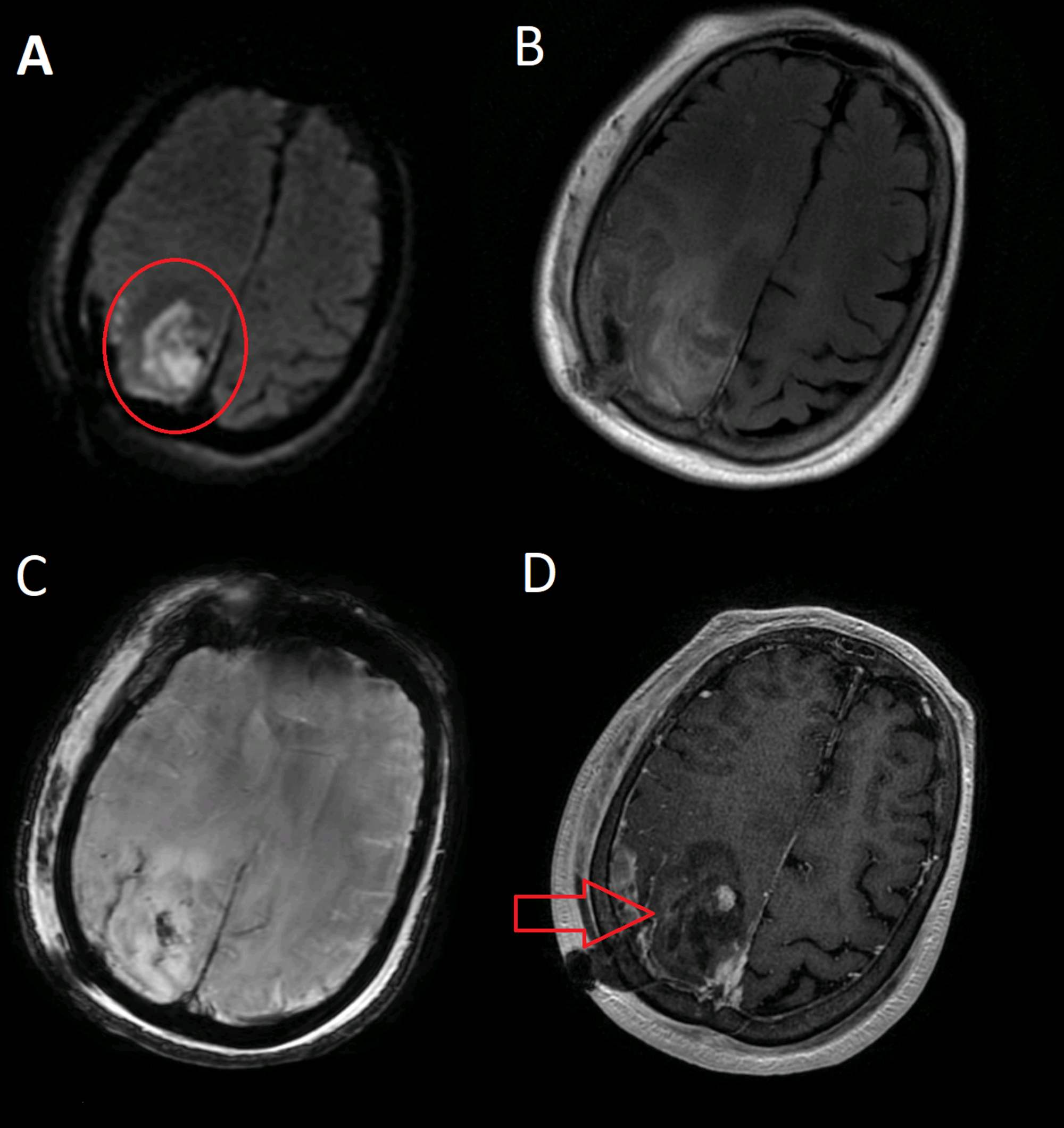
รวมกัน ภาพถ่ายเกี่ยวกับct brain contrast vs non contrast คือ ที่เว็บไซต์ vttn.vn รวบรวมและจัดทำอย่างครบถ้วนค่ะ มีภาพถ่ายที่เกี่ยวข้องกับ ct brain contrast vs non contrast คือ, contrast vs non contrast ct brain, ct brain non contrast, ct brain with cm คือ, ct brain without contrast definition, non contrast vs contrast ct, contrast vs non contrast ct head, ct brain nc คือ ที่คุณสามารถดูรายละเอียดเพิ่มเติมที่ด้านล่างค่ะ
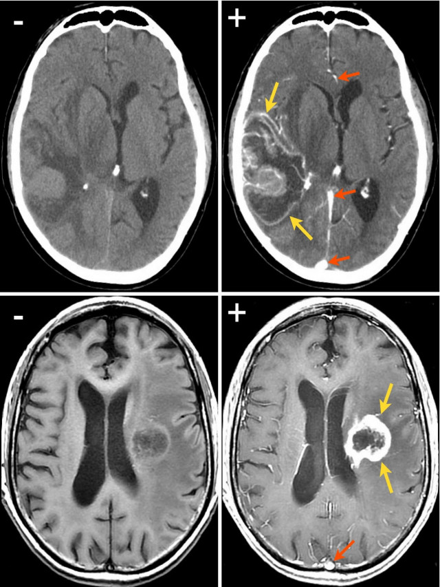
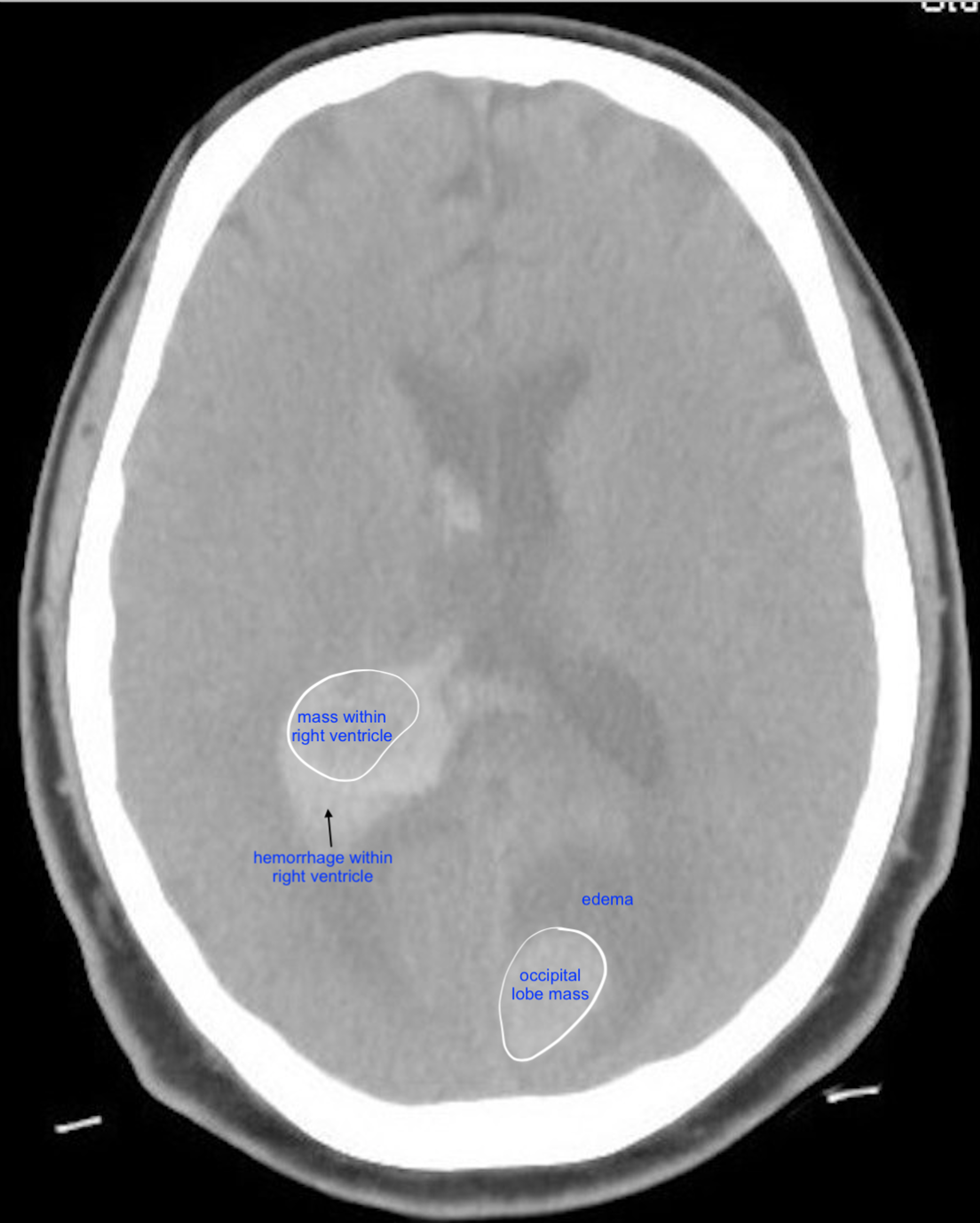
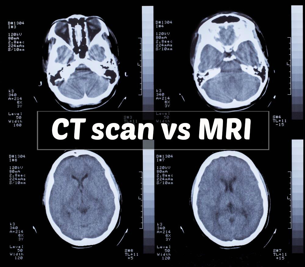
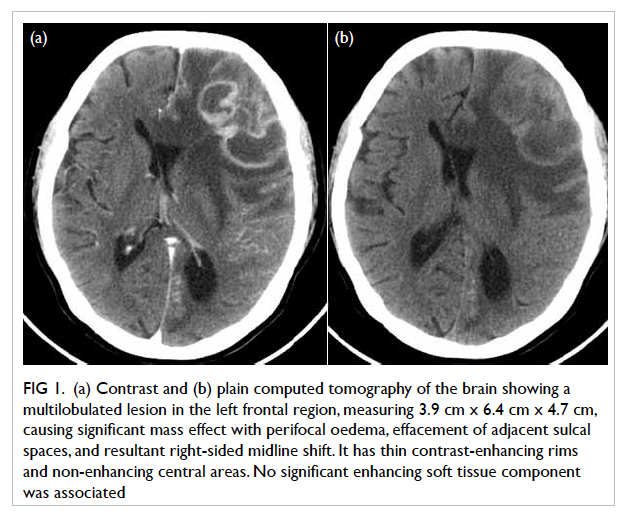
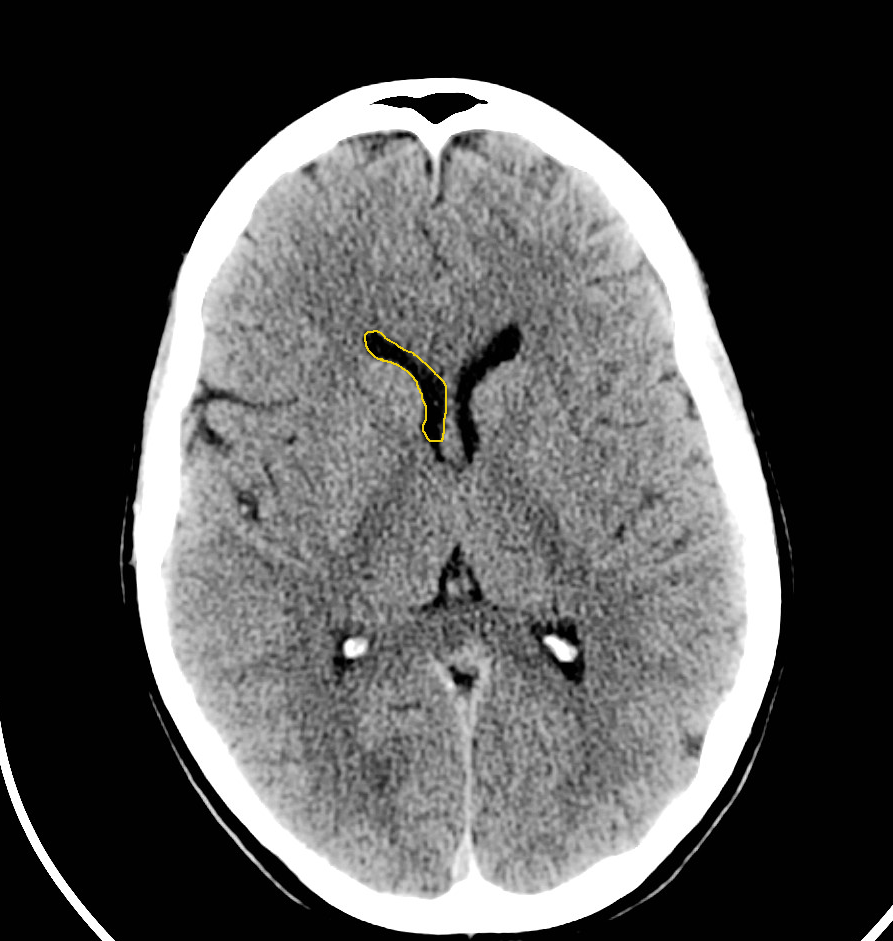
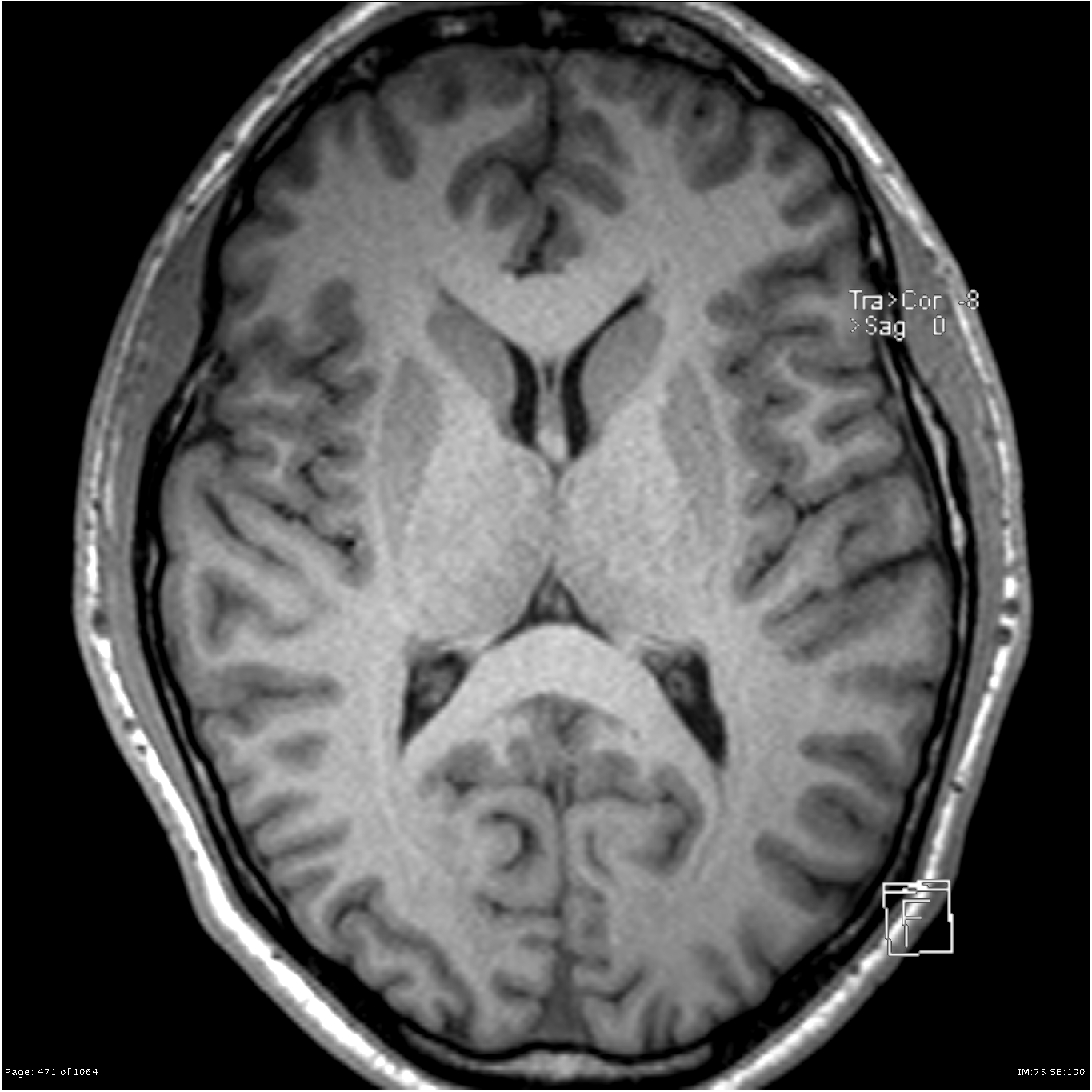
ct brain contrast vs non contrast คือ


ขอบคุณที่ต้องการเสริมสร้างความรู้ด้วยการอ่านบทความ ct brain contrast vs non contrast คือ ที่มีให้บริการที่ vttn.vn ค่ะ ท่านสามารถแสดงความคิดเห็นและตรวจสอบบทความที่เกี่ยวข้องเพิ่มเติมที่ด้านล่างค่ะ หวังว่าจะเป็นประโยชน์ในการให้ข้อมูลที่น่าสนใจให้กับท่านค่ะ
Posts: ct brain contrast vs non contrast คือ
Categories: รายการรูปภาพ
Author: vttn.vn